


Disclaimer: The following educational page was done by our own research in our own paraphrased words. Please click on each photo to view further information, resources, and citations. This is in no way professional or medical advice, but a resource to help get others started in their own research.


Achondroplasia
Achondroplasia is a genetic condition
where cartilage turns into bone at a slower rate during development. This genetic condition is caused by a change in the fibroblast growth factor receptor 3 gene. Shortened limbs of the arms and legs and curvature of the lower spine are usually a result of the ossification. The word achondroplasia means “without cartilage formation.” Achondroplasia occurs in about 1 in every 40,000 births. It is the most common form of skeletal dysplasia.


Amniotic Band Syndrome
Amniotic Band Syndrome:
The causes and underlying mechanisms that cause amniotic band syndrome are complex and controversial. Several different theories have been proposed to explain the complex mechanisms that underlie amniotic band syndrome. The two main theories are known as the extrinsic theory and the intrinsic theory. The extrinsic theory states that amniotic band syndrome occurs due to factors found outside of the fetus (externally); the intrinsic theory states that amniotic band syndrome occurs due to factors found within the fetus (internally). [see link from rarediseases.org for further breakdown of ABS theories]
In other words: Before the baby is born the body parts that show signs of ABS are entangled in string-like bands. If a band is tight enough, the constriction may cause an amputation before birth. Other times, the amniotic band may only cause a small dent around a finger or limb. Deeper bands can cause serious swelling, cut off the flow of blood, or keep that part of the body from growing in the typical way. This includes hands, feet, fingers, toes, etc.


Clubfoot
Clubfoot: True clubfoot is characterized by abnormal bone formation in the foot. There are four variations of clubfoot, including talipes varus, talipes valgus, talipes equines, and talipes calcaneus. In talipes varus, the most common form of clubfoot, the foot generally turns inward so that the leg and foot look somewhat like the letter J. In talipes valgus, the foot rotates outward like the letter L. In talipes equinus, the foot points downward, similar to that of a toe dancer. In talipes calcaneus, the foot points upward, with the heel pointing down.
To prevent future complications with walking, Orthopedic surgeons work with parents to straighten and strengthen the muscles in the foot within the first few weeks after the baby is born. The Ponseti method (which involves casting and a special boot to hold the foot in place) or French method (which consists of stretching, massages, taping and splinting) are used, leaving surgery as a last resort.


Congenital Amputation
Congenital amputation is a condition where a person is born without a limb or portion of a limb. About 1 in 2,000 babies are born with congenital amputation each year.
The condition is often caused by amniotic band syndrome, which occurs when a free floating piece of amniotic sac tissue becomes entangled with a baby’s developing limb, cutting-off the blood supply and halting development. Amnion ruptures, a condition where the internal amniotic sac becomes separated from the outer portion, contributes to free-floating tissues. However, the exact causes of most cases cannot be exactly pinpointed.
Children with congenital amputation are usually not affected by any other disabilities and are often very resourceful in compensating for their underdeveloped limb, with many being able to fully participate in most activities.


Ectrodactyly
Ectrodactyly, which is also known as split hand/foot malformation (SHFM), is a condition characterized by absence or malformation of one or more of the fingers or toes. Usually, the middle fingers or toes are affected. All four hands and feet may be affected in some individuals. However, some individuals have only mild malformation or are unaffected. Individuals with EEC may also exhibit webbing or fusion (syndactyly) of some of the fingers and/or toes. In some cases, syndactyly is the only limb defect that occurs.


Fibular Hemimelia
Fibular Hemimelia occurs when a baby is born with either part of the fibula or sometimes without it. The fibula is the thinner bone that appears between the knee and ankle. This happens more often in the right leg than left. Fibular hemimelia looks different with every person.
For some people, their leg is typically developed with only part of the fibula bone; for others their leg is curved with bowing at the knee joint and ankle.
“Here are some of the things that parents might see when a baby is born with hemimelia:
• When all or some of the bone is missing in one leg, the leg is shorter than the other. Doctors call this a leg length discrepancy.
• Because the shinbone is short or missing, the ankle joint may not form as it should. The ankle and foot might look different from normal.
• The child's knee and lower leg might bend inward.
• The child's lower leg may bow out.
• The foot may not have all five toes.”-Kids Health


Phocomelia
Phocomelia is a rare birth difference that can affect the upper and/or lower limbs. The bones of the affected limb are either missing or underdeveloped. The limb is shortened and in some cases, the hand or foot may be attached directly to the trunk. Phocomelia can be responsible for the absence of pelvic and thigh bones. Fingers and toes may also be fused together. While the most common symptom of phocomelia is the malformation of the limbs, differences of the eyes, nose, and ears may also be present. If all four limbs are absent, it’s called tetraphocomelia. “Tetra” means four, “phoco” means seal, and “melos” means limb. This term refers to the ways the hands and feet look. The hands may be attached to the shoulders, while the feet may be attached to the pelvis.
Upper and lower extremity prosthetics are available for legs and arms as a result of Phocomelia. Infants with Phocomelia can be introduced to artificial limbs as early as 6 months to begin adapting to life with a prosthetic if desired.


Proximal Femoral Focal Deficiency
Proximal Femoral Focal Deficiency (PFFD) is a rare condition affecting about 1 in every 200,000 births, where the upper part of the femur (thigh bone) is either missing or malformed. This causes one leg to be shorter than the other and can cause functional issues with a child’s ability to walk. Malrotation, limb-length discrepancies, and fibular hemimelia can be associated with PFFD. There are several treatment strategies and alternatives for PFFD such as prosthetics, corrective surgery, and leg lengthening or modification procedures.


Radial Dysplasia
Part 1. Radial longitudinal deficiency is a rare condition that affects the forearm. It is a congenital condition (not inherited) and can affect one or both arms. It is sometimes called radial club hand, radial dysplasia, or radius deficiency.
There are two bones in your baby’s forearm: the ulna, on the outer side of the arm, and the radius, on the inner side. Radial longitudinal deficiency occurs when the radius does not form properly. This causes the wrist to bend toward the thumb side of the forearm.
Radial longitudinal deficiency also affects the soft tissues and flesh of the forearm. The arrangement of muscles and nerves may be unbalanced, and some muscles and nerves may be missing-BCH
RLD encompasses a spectrum of complex congenital malformations of the radial side of the forearm. These are uncommon and can range from a mildly hypoplastic thumb to a completely absent radius. Radial club hand is commonly associated with a number of syndromes for which evaluation must occur. Mild deficiency can be managed nonoperatively, whereas severe cases are usually treated with surgery.

Radial Dysplasia 2/2
Part 2. Radial longitudinal deficiency/Radial Dysplasia/Radial Club Hand
There are four types of radial dysplasia dysplasia. Signs and symptoms depend on what type of radial dysplasia a child has.
Type I
This is the mildest form of radial dysplasia. The radius is just a little shorter than normal and the wrist turns in only slightly.
Type II
The radius is much smaller than usual and the wrist is more turned in.
Type III
A large part of the radius missing and there is more severe turning in of the wrist. The other bone of the forearm (the ulna) is curved and thickened.
Type IV
There is no radius at all. The wrist is very turned in.
In all types, the thumb may be smaller than usual or completely missing. Radial dysplasia can happen on one or both sides.
Radial dysplasia happens while a baby is developing in the womb. It may be part of a syndrome syndrome known as VACTERL syndrome.
• Vertebral (spine) differences
• Anal atresia (an anus that does not open to the outside of the body)
• Cardiac (heart) problems
• Tracheo-Esophageal fistula fistula(a connection between the breathing and swallowing tubes)
• Renal (kidney) issues
• Limb differences in addition to the radial dysplasia
Sometimes it can happen as part of other genetic syndromes where there are other underlying medical issues. -Kids Health


Symbrachydactyly
Symbrachydactyly has been subdivided by Blauth and Gekeler (see NIH study) into 4 types:
* (1) Short finger (brachymesophalangia): all 4 fingers are short and incompletely formed with a relatively normal thumb; middle phalanges may be missing with incomplete syndactyly.
(2) Oligodactylic (atypical cleft hand): the central portion of the hand is aplastic, whereas the borders digits are less severely affected.
(3) Monodactylic: the fingers are absent or aplastic, whereas the thumb is present.
(4) Peromelic: resembles a transverse amputation at the metacarpophalangeal joint level, may have nubbins with or without nail remnants.
In most cases, children born with symbrachydactyly are able to adapt to their physical limitations and experience a fully functional life with no treatment.


TARS
TARS: Thrombocytopenia Absent Radius Syndrome (TARS) is a rare genetic condition where children are born without radius bone (the bone that forms right behind the thumb into the fold of the elbow) often children with TARS have a low platelet count (the blood cells that play a huge role in clotting). Children with TARS usually have all 5 fingers - just not the Radius bone. Children with TARS may need blood transfusions to help stabilize their platelet count. Often kids with TARS have a little dimple in their wrist - this picture captures that. They are often sensitive to dairy, reflux is very common.


Tibial Hemimelia
Tibial Hemimelia (also known as tibial deficiency) is a condition in which a child is born with a tibia (shinbone) that is shorter than normal or missing altogether. This creates a difference in the length of the child’s legs. The condition is extremely rare, occurring in only about one out of every one million births. There are about four new cases in the United States each year.
The condition may be bilateral in about 30% of patients and associated with other anomalies, the most common of which is deformities of the foot, although deformities of the ipsilateral femur, hip dysplasia, upper extremity and spinal anomalies have also been reported. Sometimes, Tibial Hemimelia runs in a family or is part of a medical condition. Most of the time, however, doctors are not able to determine exactly why the Tibia is shorter.
Almost all children with Tibial Hemimelia will need surgery to help them stand, walk and play better. The type of surgery needed depends on several different factors, including how much of the tibia is present and the condition and stability of the child’s knee and ankle joints.


Ulnar Longitudinal Deficiency
Ulnar longitudinal deficiency, previously known as ulnar club hand, is a condition in which your child’s wrist is in a fixed and bent position toward the side of the hand with the little finger. This condition is also sometimes called ulnar dysplasia. It happens when 1 of the long bones of the forearm, the ulna, and other soft tissues of the hand, develop differently in the womb. Your child’s fingers and thumb may also be affected. Other muscles and nerves in the hand may be unbalanced or missing.
Children may also have other malformations in the musculoskeletal system, such as scoliosis.
Ulnar longitudinal deficiency affects about 1 in 100,000 babies.
Infants with ulnar longitudinal deficiency can wear a splint and be treated with gentle stretching exercises to help their wrist and elbow move into a normal position and recover some range of motion. Some types of ulnar longitudinal deficiency can also be treated with surgery to improve hand and arm function.


Ulnar Mammory Syndrome
Ulnar Mammory Syndrome or Schinzel Syndrome.
The main features of UMS include upper limb defects (including abnormal or incomplete development of the fingers and forearm), underdevelopment of the mammary and apocrine glands (leading to absent breast development and the inability to produce breast milk), and various genital abnormalities. Other signs and symptoms may include hormonal deficiencies.
UMS is caused by mutations in the TBX3 gene and inheritance is autosomal dominant. However, not all people who have or inherit a mutation will have features of UMS. This phenomenon is called incomplete penetrance. Treatment depends on the specific symptoms and severity in each person and may include surgery to improve the function or appearance of limbs, and hormone replacement therapy if hormonal deficiencies are present.
The exact prevalence of UMS is not currently known, but less than 150 cases have been reported in the medical literature to date.


VACTERL
VACTERL association is a nonrandom association of birth defects that affects multiple anatomical structures. The term VACTERL is an acronym with each letter representing the first letter of one of the more common findings seen in affected children:
(V) = vertebral abnormalities
(A) = anal atresia
(C) = cardiac defects
(TE) = tracheal-esophageal abnormalities, including atresia, stenosis and fistula
(R) = renal and radial abnormalities
(L) = limb abnormalities
Babies who have been diagnosed as having VACTERL association usually have at least three or more of these individual anomalies. The syndrome has been estimated to occur in one in 10,000 to 40,000 newborns.
Limb differences occur in up to 70 percent of babies with VACTERL association. These differences may include radial aplasia, radial hypoplasia, and triphalangeal thumb. Other limb anomalies including polydactyly, syndactyly, radioulnar synostosis and lower limb malformations such as clubfoot, and hypoplasia have been described in VACTERL association.
Babies with limb differences on both sides tend to have kidney or urologic defects on both sides, while babies with limb defects on only one side of the body tend to have kidney problems on that same side.


Sepsis Amputation
“Sepsis is a condition that occurs when the body’s response to an infection damages its own tissues and can lead to extensive inflammation, blood clotting, and organ failure. As sepsis worsens, sometimes nutrients cannot get to the tissues in the fingers, hands, arms, toes, feet, and legs. The body’s tissues begin to die and can cause gangrene. The dead tissue must be removed to prevent further infection. If the damage is extensive, amputation may be needed. Out of the approximate 185,000 amputations per year, around 13,870 are caused by sepsis.
On average, about 38 amputations a day are due to sepsis. Amputation is the loss or removal of a body part. The decision to amputate is based on making sure enough tissue is removed to ensure that all the damaged tissue is gone, and to preserve the patient’s independence and mobility as much as possible. Ideally, patients who have had an amputation will begin physiotherapy as soon as possible. Physiotherapy may include stretching and increasing muscle strength. Occupational therapists play an important role in helping amputees learn how to use special tools or adapt their living environment as needed. ”

Apert Syndrome
“Apert syndrome also known as acrocephalosyndactyly, is a genetic difference that causes fusion of the skull, hands, and feet bones. Apert syndrome occurs in one out of every 65,000 to 88,000 births.”
Children with Apert syndrome typically experience craniosynostosis and midface hypoplasia. The characteristic feature that distinguishes Apert syndrome from other types of syndromic craniosynostosis is the presence of syndactyly— a condition in which their fingers are webbed or conjoined. There are three main types of syndactyly in Apert syndrome: Type I (“spade hand”): the index, middle, and ring fingers are fused, and the thumb is free. These patients typically have a “flat” palm.
Type II (“mitten hand”): the three middle fingers and the thumb are fused, and the palm curves inward or is concave. Type III (“rosebud hand”): All fingers and the thumb are fused.
Complex syndactyly benefits from specialty surgical care to release and result in the greatest degree of function that can be achieved.”


Tibial Torsion
Tibial Torsion
tib·i·al | \ ˈtib-ē-əl : of, relating to, or located near a tibia. \ tor·sion | \ ˈtȯr-shən : the twisting of a bodily organ or part on its own axis.
There are two types of tibial torsion: internal & external. Whether the toes point inward (intoeing) or outward (outtoeing), tibial torsion is a common cause of gait (manner of walking) differences. Other conditions that can cause intoeing include:
• femoral anteversion, the inward twisting of the thigh bone (femur)
• metatarsus adductus, a foot deformity that causes the front half of the foot to turn inward.
Almost all children with internal tibial torsion improve without treatment, usually by the time they turn 4.
Although tibial torsion is a limb difference that you’re born with, it is often unnoticeable until a child starts to walk. For many kids, one leg twists more noticeably than the other.

Camptodactyly
“Camptodactyly is a rare congenital condition, affecting about 1% of the population, characterized by a digital flexion deformity that occurs in the proximal interphalangeal (PIP) joint. This can be caused by an underlying abnormal structure of the finger, including contracted tendons or ligaments, irregularly shaped bones, and tight skin. Camptodactyly can present at birth as one or multiple fingers are bent, though can be more noticeable throughout childhood. Sometimes, individuals have a family history, or it can be part of a syndrome that impacts multiple parts of the body. Milder presentations may not cause symptoms aside from the finger being slightly curved. More prominent and severe cases can affect hand function. Treatment depends on the severity and the underlying cause of the condition. The treatment typically involves observation with a passive stretching regimen. However, surgical intervention may be an option for certain cases.”

Holt Oram Syndrome
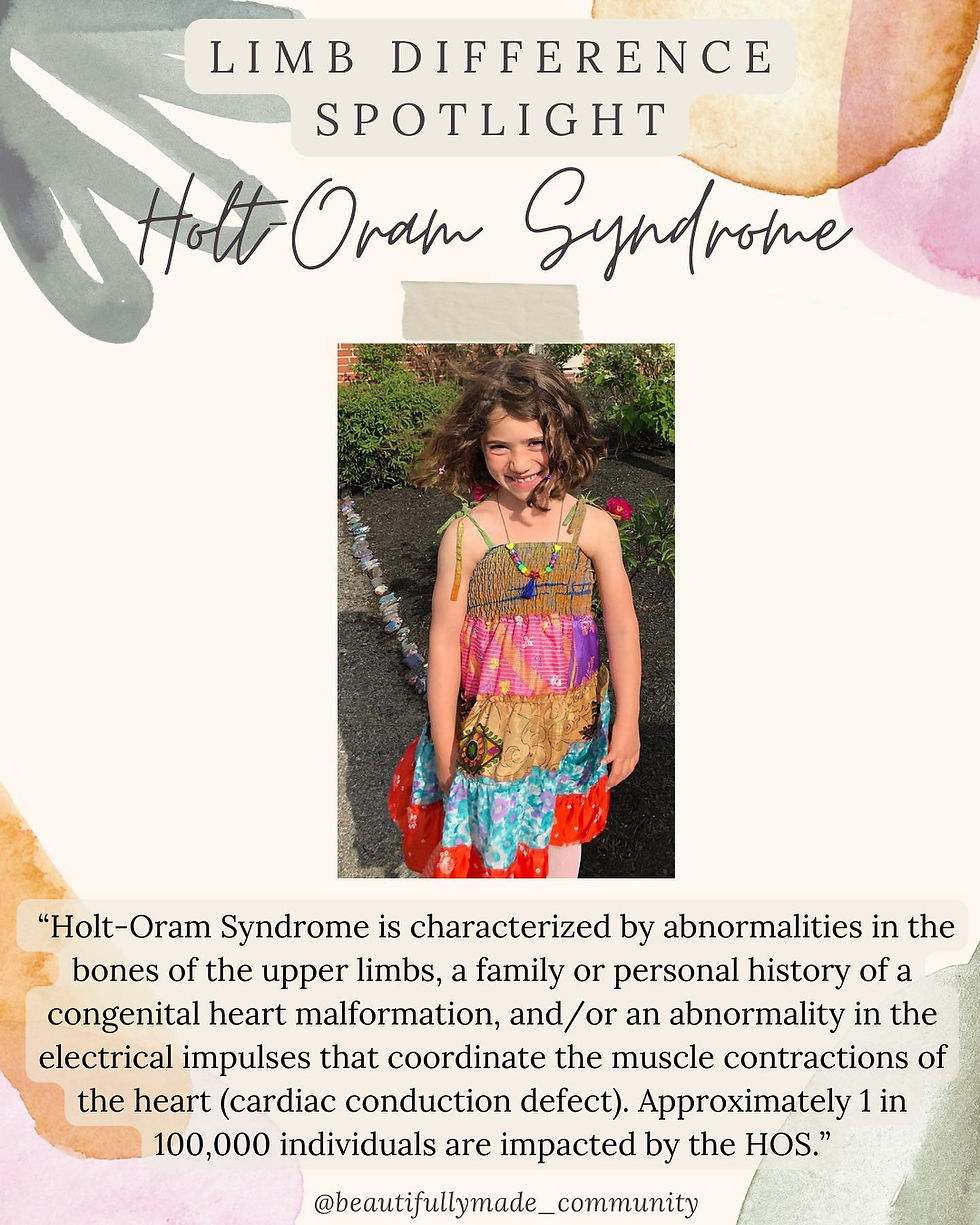
“Holt-Oram Syndrome is characterized by abnormalities in the bones of the upper limbs, a family or personal history of a congenital heart malformation, and/or an abnormality in the electrical impulses that coordinate the muscle contractions of the heart. HOS is an autosomal dominant genetic condition that is associated with a change in the TBX5 gene. Most cases result from a new mutation in patients without a family history of the disorder. In some individuals, an abnormal wrist bone is the only evidence of the condition. Individuals with HOS may have additional bone abnormalities including a missing thumb, a long thumb that looks like a finger, partial or complete absence of bones in the forearm, an underdeveloped bone of the upper arm, or abnormalities of the collar bone or shoulder blades. 75% of individuals also have a congenital heart condition. The most common is a defect in the muscular wall that separates the right and left sides of the heart. A hole in the septum between the upper chambers of the heart is called an atrial septal defect, and a hole between the lower chambers of the heart is called a ventricular septal defect. Some people have cardiac conduction disease, which is caused by abnormalities in the electrical system that coordinate contractions of the heart. Approximately 1 in 100,000 individuals have the Syndrome.”


Posteromedial Tibial Bowing
“Posteromedial Tibial Bowing is a congenital condition, impacting around 1 in 40,000, in which there is both posterior and medial bowing of the tibia and fibula. The foot assumes a calcaneovalgus position and is typically in extreme dorsiflexion. The foot at birth will occasionally be abutting the concave tibia and a skin dimple is often present over the apex of the bow. The bowing may partially correct on its own within the first 4 years of life. Although the tibia's growth will be inhibited, treatment is always individualized to accommodate predicted height and the difference in limb lengths.”

Tibial Torsion
Tibial Torsion
tib·i·al | \ ˈtib-ē-əl : of, relating to, or located near a tibia. \ tor·sion | \ ˈtȯr-shən : the twisting of a bodily organ or part on its own axis.
There are two types of tibial torsion: internal & external. Whether the toes point inward (intoeing) or outward (outtoeing), tibial torsion is a common cause of gait (manner of walking) differences. Other conditions that can cause intoeing include:
• femoral anteversion, the inward twisting of the thigh bone (femur)
• metatarsus adductus, a foot deformity that causes the front half of the foot to turn inward.
Almost all children with internal tibial torsion improve without treatment, usually by the time they turn 4.
Although tibial torsion is a limb difference that you’re born with, it is often unnoticeable until a child starts to walk. For many kids, one leg twists more noticeably than the other.


Femoral Anteversion
“Femoral anteversion is a condition where the femur (thigh bone) twists inward. The inward twisting of the femur causes the knees and feet to turn in, or have a ‘pigeon-toed’ appearance. Femoral anteversion occurs in approximately 10% of children, and the cause is unclear. Femoral anteversion often resolves on its own as the child grows and does not necessitate treatment. In more severe cases that do not self-correct by age 8 or 9 years old, treatment may involve bracing and special shoes aimed at correcting the foot position, or surgery to rotate the femur to a better position.”